Heart and Lungs
Structure and Function of the Heart
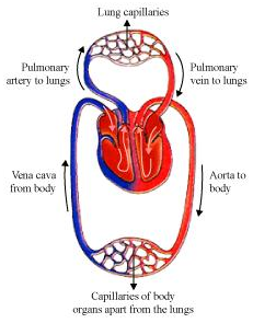
The heart has 4 chambers (Right and left ventricle, right and left atrium), and it is a muscular pump that circulates blood around the body. The right side of the heart only pumps de-oxygenated blood towards the lungs so that it can pick up oxygen. The left side of the heart only pumps oxygenated blood from the lungs to the rest of the body. The heart is extremely important in transporting substances such as food, water, oxygen, waste products around the body. Humans have a double circulatory system, as deoxygenated blood has to be pumped through the lungs to become oxygenated, before it can return to the heart so that the heart can pump the oxygenated blood around the body. The natural resting heart rate is controlled by a group of cells located in the right atrium that act as a pacemaker. Artificial pacemakers are electrical devices used to correct irregularities in the heart rate.
Deoxygenated blood enters the right atrium through the vena cava → The blood is then pumped through a valve and into the right ventricle chamber → Blood goes up through the pulmonary valve, and into the pulmonary artery (which goes towards the lungs) → the blood is now oxygenated, and enters the pulmonary veins and into the left atrium → Blood is then pumped through a valve, and into the left ventricle → Blood then flows through the aortic valve, and out of the aorta towards the rest of the body. (NOTE: Right and left are reversed when we talk about the heart. E.g. you’ll see that the right ventricle is on the left side of the heart.

The heart has a special type of muscle that is extremely thick, called the cardiac muscle. This is because the muscle is constantly contracting and relaxing, therefore it has to be quite tough. The muscle has its own blood supply, through the coronary artery. This provides the heart with oxygen and glucose in order to function. The heart has important valves to prevent the backward flow of blood, as deoxygenated blood is flows quite slow and with a low pressure.
Structure and Function of the Lungs
| Part of the Lung | Function |
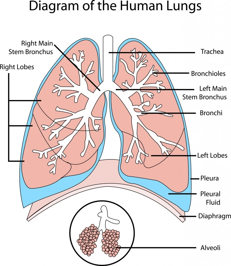
| Trachea (windpipe) | Where the air enters, and it leads to the bronchi |
| Bronchi | Bronchi then splits off into two so that there is a bronchi leading to the left lung, and a bronchi leading to the right lung. The bronchi then branches off into bronchioles |
| Alveoli | The bronchioles then lead to the alveoli, which are microscopic sacs where gas exchange takes place |
| Capillary Network surrounding the alveoli | Extremely thin capillary network around the alveoli so that there is less of a distance for the gas to diffuse across |
Alveoli Adaptation
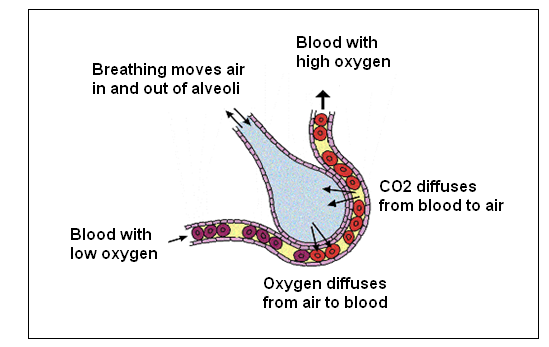
Alveoli are highly adapted for gas exchange by diffusion between air in the lungs, and blood in the capillaries. They are folded in order to maximise the surface area to volume ratio. This allows more gas exchange to take place. The alveoli wall linings are moist, allowing oxygen and carbon dioxide to dissolve easily. It has thin walls so that the distance in which the gases have to diffuse across is short. Capillaries have a rich blood supply, which intensifies the concentration gradient.
- Which is the part of the lungs where the air enters?
- Trachea
- How many chambers are there in the heart?
- Your answer should include: Four / 4