The Respiratory System
Cell Organisation

Cells
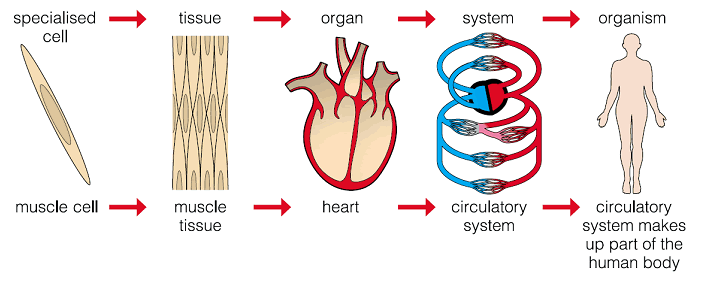
Cells are the building blocks of life. When cells become specialised they can carry out a particular function. The process of a cell becoming specialised is called cell differentiation. Cells need to be specialised in order to form multicellular organisms.
Tissues
Specialised cells come together to form tissues. A tissue is a group of cells that work together towards a specific function. Lots of animals have more than one type of tissue (for example, skin tissue, muscular tissue, glandular tissue).
Organs
An organ is a group of tissues that work together towards a particular function. For example, the stomach is an organ which is made out of muscle tissue, epithelial tissue and glandular tissue - all working together to break down food.
Organ system
An organ system is a group of organs that work together towards a particular function. For example, the digestive system is composed of lots of organs: stomach, liver, glands, small intestine and large intestine.
Living Organism
A living organism is a group of organ systems that work together to form a living creature.
__A living organism is a living creature that abides by the MRS GREN rules. __
__For something to be living, it must have the ability to: __
- Move
- Respire
- Sense
- Grow
- Reproduce
- Excrete
- Nutrition
The Lungs
Not all living organisms have lungs, but since humans have lungs, it’s important to learn about them! Human cells need oxygen in order to respire.
Respiration is a chemical process in which energy is produced from reacting oxygen and glucose:

The lungs are responsible for getting Oxygen into the body for respiration!
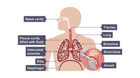
The lungs are in the thorax (top part of your body) and protected by the ribcage. The diaphragm is a large slab of muscle which lies underneath your lungs.
Here is the path air takes from outside of the body to the bloodstream:
Mouth - Trachea - Bronchi - Bronchiole - Alveoli - Capillary
Trachea
The trachea is a long tube connecting your mouth and bronchi. There are rings of cartilage throughout the trachea to stop the airway from closing.
Bronchi
The bronchus (plural, bronchi) is a smaller tube which leads from the trachea into the lung. There are two lungs and a bronchus leads into each one.
Bronchioles
Each Bronchus splits into hundreds of smaller tubes, called bronchioles.
Alveoli
Each bronchiole leads into tiny air sacs called an alveoli (singular, alveolus).
Capillary
Each alveoli is surrounded by at least 1 capillary. A capillary is a tiny blood vessel.
Alveoli
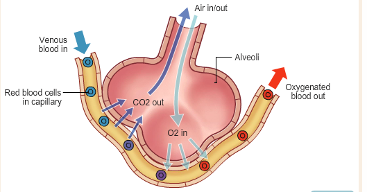
When you breathe in, oxygen rich air enters an alveoli. The oxygen absorbs through the walls of the alveoli and onto a red blood cell within the capillary. In the meantime, the passing unoxygenated blood releases waste into the alveoli, ready for you to breathe it out.
You will need to know the 4 adaptations of the alveoli:
- They have a large surface area for efficient gas exchange - since they are small and there are thousands of them.
- Their walls are only 1 cell thick - so that gas can easily absorb through the walls into the capillaries.
- Their walls are moist - so that gas can easily absorb through the walls.
- They have a good blood supply - so that gas exchange is quicker and more efficient.
Double Circulatory System
The circulatory system is the transport system within the body which transfers oxygen and food around the body (as well as picking up waste and getting rid of it).
Humans have a double circulatory system.
The heart plays a big part in the circulatory system, so we will learn more about that in the next section. Let’s take a journey through the circulatory system of a red blood cell, starting at capillary passing an alveoli.
- The red blood cell picks up oxygen from the alveoli
- The oxygenated red blood cell travels through the blood vessels towards the left side of the heart.
- The heart pumps the blood and the red blood cell is pumped through the blood vessels towards the body cells
- When the red blood cell passes a body cell that needs oxygen, the oxygen is released and enters the body cell.
- The now unoxygenated red blood cell travels towards the right side of the heart
- The heart pumps the blood and it travels quickly towards the lungs
- The red blood cell picks up oxygen from the alveoli - and the whole cycle starts again!!
It is called a double circulatory system since there are in effect two circuits of the blood.
The Heart
The heart is made up of four sections:
- A left atrium
- A right atrium
- A left ventricle
- A right ventricle
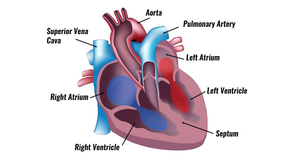
An easy way to remember which section is which is that the atriums are at the top and the ventricles are on the bottom. The left atrium and left ventricle are on the right side of the picture. This is because it is as if you are looking at someone else’s heart in from of you!
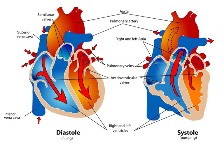
Movement of blood
The heart’s main function is to pump blood around the body. This is split into 4 chambers in order to keep the oxygenated blood and the deoxygenated blood separated.
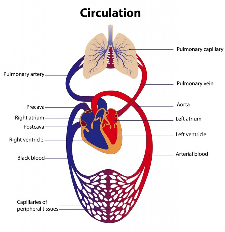
- Blood flows into both the left and right atrium from the vena cava and pulmonary vein.
- The two atria contract, pushing the blood through the valves and into the ventricles.
- The two ventricles contract, pushing the blood through the next set of valves and out through the pulmonary vein and aorta.
- Blood in the aorta travels around the rest of the body, providing cells with oxygen. The blood then travels back through to the vena cava.
- Blood from the pulmonary artery travels to the lungs to be oxygenated. The blood then travels back to the pulmonary vein.
The cycle starts again!
Valves
Valves stop the blood from flowing backwards.
The heart has 4 valves:
- One separating the left atrium and left ventricle
- One separating the right atrium and right ventricle
- One separating the pulmonary artery and right ventricle
- One separating the aorta and left ventricle.
Pacemakers
Everyone is born with a natural pacemaker. The walls of the right atrium contain cells which release electrical signals through the heart, causing the muscles to contract at the right time!
If someone’s heart has stopped beating properly (or is struggling to beat properly) then an artificial pacemaker can be put in. This is placed under the skin and has an electrical wire going to the heart.
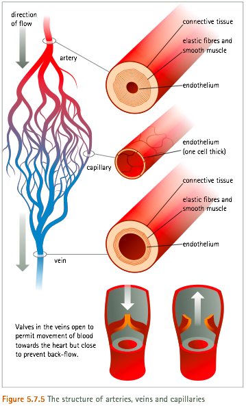
The Blood Vessels
Arteries
- Carry blood away from the heart
- Have thick, strong and elastic walls - since blood coming from the heart is at high pressures (since it has just had a big pump from the heart).
- The elastic fibres help them to spring back
- Walls are thick compared to the lumen (hole in which blood flows).
Capillaries
- Arteries branch into lots of small capillaries
- Capillary walls are 1 cell thick - for easy substance exchange.
- Capillaries are used for material exchange - such as diffusion of oxygen into cells.
- Very small and narrow - so the surface area to volume ratio is very high (for efficient material exchange)
Veins
- Capillaries join up to form veins
- Blood is at a lower pressure in veins (since it has slowed down).
- Veins have a large lumen compared to the walls of the vein - to allow a good flow, despite the low blood pressure.
- The walls are not as thick or strong as arteries (since they aren’t dealing with high pressures).
- Veins contain valves, which prevents back flow.
The Blood
What is blood
- Blood is a group of similar cells which work together to transport substances around the body. So… Blood is a tissue!
- Blood is composed of red blood cells, white blood cells, platelets and plasma.
- Waste material (such as urea and carbon dioxide) and useful materials (such as oxygen and glucose) are also found in the blood, but these are not part of blood. Think of blood as a river and the river can carry things along with it!
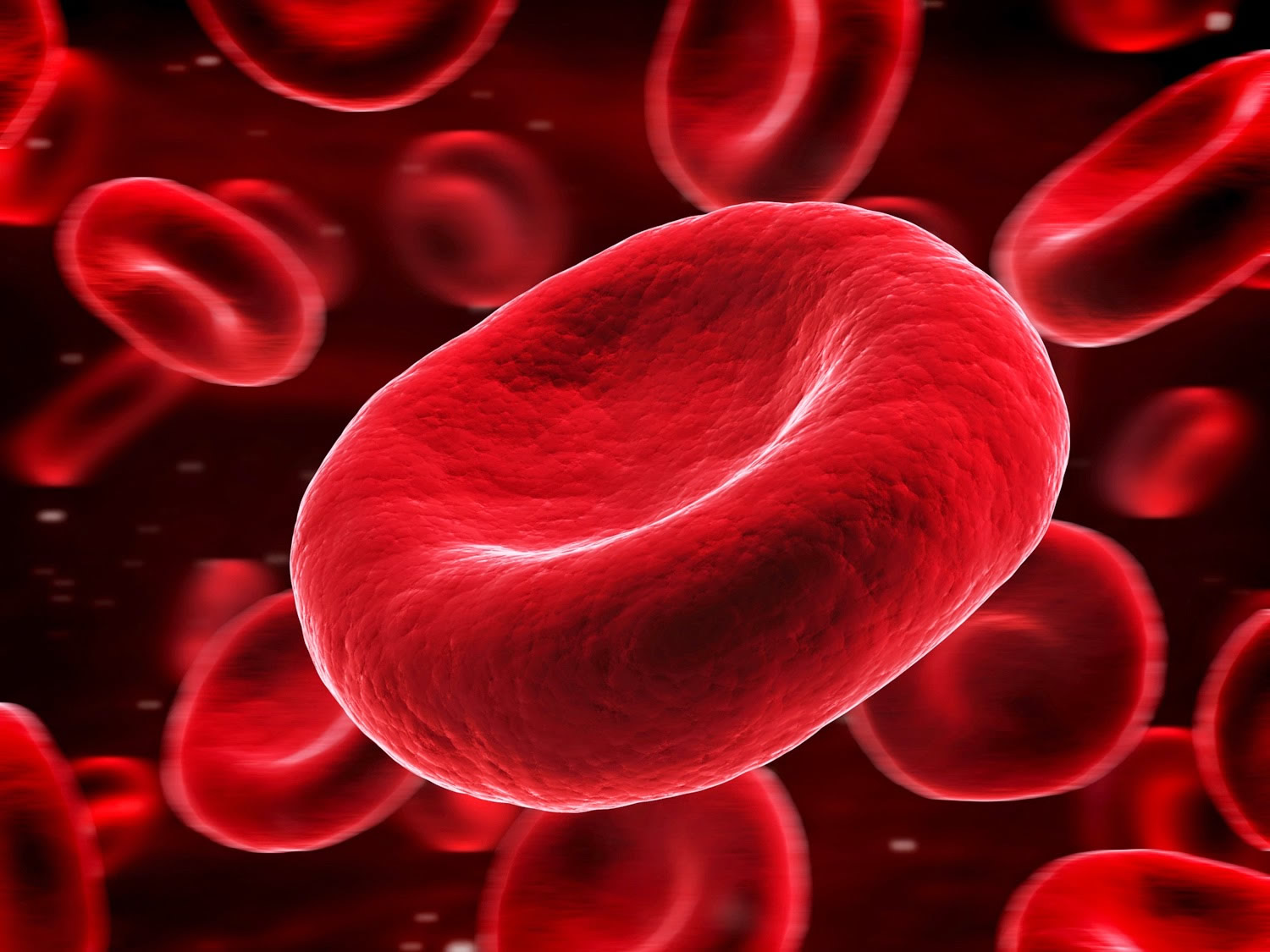
Red blood cells
Function: Carry oxygen around the body.
- They have a biconcave structure - to give them more surface area to carry more oxygen
- Contain a red pigment called haemoglobin - this actually binds to the oxygen.
- Doesn’t have a nucleus - allows more room for oxygen
- When haemoglobin is combined with oxygen it is called oxyhaemoglovin
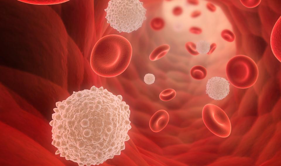
White blood cells
Function: to defend the body against disease causing microorganisms.
- Different types of white blood cell that protect you in different ways
- They can engulf and digest harmful microorganisms
- They can produce antibodies that fight harmful microorganisms
- They can produce antitoxins that neutralise any toxins that are released by harmful microorganisms
- White blood cells do have a nucleus.
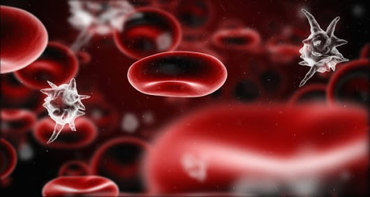
Platelets
Function: to help blood to clot (like when a scab forms over a wound) in order to stop you from losing too much blood from a wound and to prevent harmful microorganisms from entering the wound.
- Platelets are small fragments of cells
- They don’t have a nucleus
- They clump together to clot blood
- Blood clotting helps us to not lose too much blood from a wound.
- Blood clotting helps to protect microorganisms from entering a wound.
Plasma
Function: A pale yellow liquid that carries everything in the blood.
- Carries red and white blood cells and platelets
- Carries nutrients that are absorbed by the small intestine and carries them to cells.
- Carries carbon dioxide from cells to the lungs
- Carries urea from the liver to the kidneys.
- Carries hormones and proteins from place to place.
- Carries antitoxins and antibodies which are produced by the white blood cells
